So, I sorta faked you out. I wanted to blog last Friday, but ended up having to do a TON of laundry to prepare to go to Yozgat the next day. Then, arriving in Yozgat, we found no internet. We didn't get back until late Thursday night, and I worked all day yesterday. So, here we are. Another week later. I sincerely apologize for the delay.
Anyhow—
I'll start with some photos from my work since that's what I spend 85% of my time doing.
These are WAY old, just so you know.
This photo is of the fields in Eskişehir. This was my first experience with field work. I found I like working outdoors. I also found that the sun in Turkey is very intense. I was sunburnt badly this first day, but it has since progressed into a very nice (farmers) tan, so no worries. If you didn't already realize, this is a picture of wheat fields. Here it is still green. Now, it has dried, and I think the particular field was harvested.
The objective for the day was to discard (negatively select) breeds of wheat based upon their susceptibility to yellow rust, leaf rust, and/or stem rust. It is done while the wheat is green because after the wheat has dried, the presence of these rust varieties is harder to detect. We "deselected" them by breaking the stems.
(In this photo: tall man w/ the hat, Dr. Alex Morgunov; the girl in the purple shirt, Cemale Mursalova; the girl in the white lab coat, Esra Musluk)
Like this. This particular plant had a mild susceptibility to yellow rust (very hard to see). Because of the millions of plants they have sown in field testing, you should only select the very best. The rest are discarded.
This is a very good plot of wheat. It has a short height (to prevent folding), strong stem, rust resistance, large spikes, long peduncle, etc.
I think I've mentioned Fusarium sp. before. This is one of the most tell-tale symptoms: white head. As you can see, the wheat stem remains green, but the spike has prematurely dried.
This photo is the most tell-tale sign of Fusarium culmorum. This particular species is commonly known as crown rot. The area where the stalk is broken is just above the crown. The browning you see in the stalk indicates the presence of the crown rot fungus. This is, to my relatively untrained eye, mildly infected plant; however, this variety is still not wanted on account of it's susceptibility.
This is a fantastic example of yellow rust. The leaves are not drying but covered in a fine yellow dust, Puccinia sp. This cultivar is highly susceptible. Obviously, I immediately discarded it.
While we performed negative selection, Dr. Morgunov was busy with positive selection. He selected very specific wheat plots for traits they possessed. The ultimate goal is to breed a short species of wheat with high yield that's resistant to disease and water stress.
So that's the field work I started with. I have a lot more pictures, but they're not uploaded and my camera battery is charging. You can look forward to more field work later.
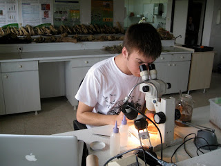
Allll right. Moving right along. Now, as I've mentioned before, I spend most of my time in the lab. I've also told you that I spend a lot of time under the microscope. I figured, then, I should take some nice microscope shots so you can see exactly what I'm working with.
This is what I dig through all day. It's a circular dish (obviously). The ridges help to keep your place and separate the soil. There's also a red line (not depicted) that helps you know where you started.
This is what I'm looking for. That lemon-shaped little... thing... is a nematode cyst. It's the mature form of a female CCN (cereal cyst nematode). It's full of nematode eggs and is very delicate. I use a pair of forceps to gently remove it from the soil and place it in sterile water.
Here's a brief bit of nematode biology:
The juveniles hatch from their eggs after a cold period. They swim (very slowly) through the wet soil until they find the root of a cereal plant. They penetrate the root and begin feeding. Eventually, they align with a specific sex. The females begin producing eggs which are fertilized by the males. The females swell. They resemble ^^above, but white. Then they die and become the brown cysts.
This is a male. This is a resembles a larger version of the vermiform juveniles (in other words, it looks similar to the wiggly little baby nematodes).
I actually just found this picture while blogging. The little guy above is a juvenile. So, there you are. This is a nice juxtaposition of the two vermiform types of nematodes.
These next two are my favorites. I think they really depict the nematodes well. You can really distinguish the stylet (the mouthpart) that it uses to penetrate the root. Here, if you can't tell, it's the upper end. The one to the right. The stylet almost appears to have a line down it. It's also very sharp.
This is under a higher-powered microscope. They are really good shots of the stylet, but the rear always seems to blur out (it's on a different plane... unfortunately at this magnification, it's nearly impossible to focus on the entire nematode well).


I've also branched out and've begun working with some experiments. This is a growth room experiment involving nematodes. I personally built all of those systems. Just sayin'.

Alright, that's enough for tonight. I'm going to attempt to get some sleep now. If you're curious why I'm up at 02:00, that would be because my face is being dive-bommed by the annoying little millers. I've killed I think nine now. More recently, the wolves have begun howling. If you recall (or if you don't, scroll down) the dogs roaming around campus. Well, they're answering the wolves. It's a fantastic chorus.... if you're not trying to sleep.
Expect more soon. I'll try to blog tomorrow.
Tekrar Çok Teşekkür Ederim
İyi geceler,
Andrew


